CHT Hydroxyapatite media
Multiple purification mode

Multiple modes
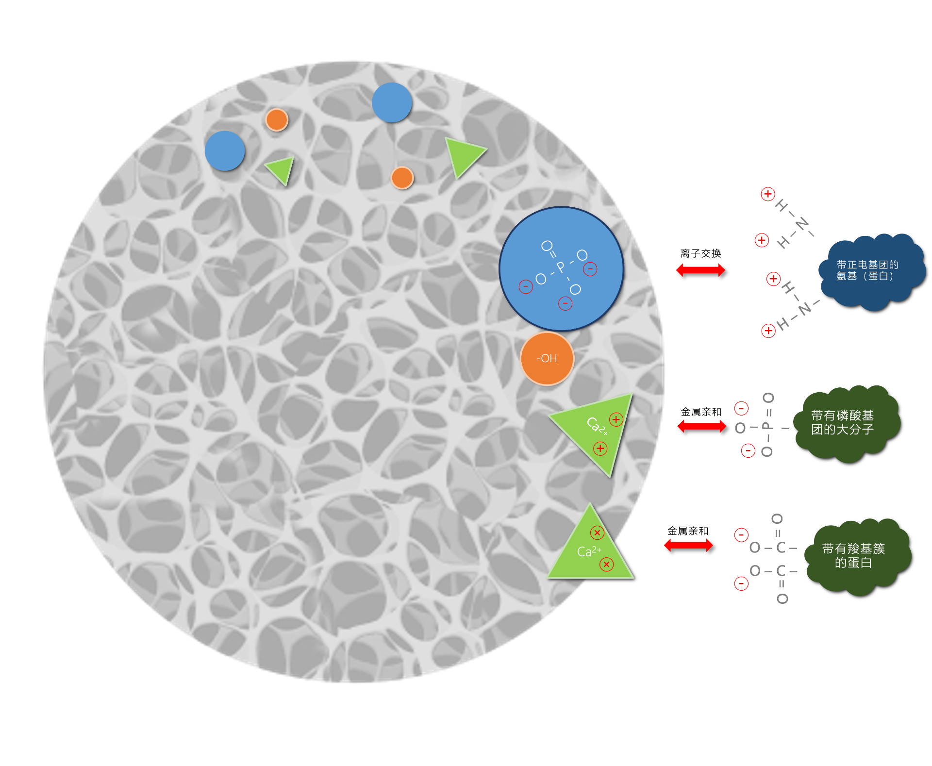
Hydroxyapatite features two distinct binding sites: Ca²⁺ and PO₄³⁻. These binding sites are distributed according to its intrinsic crystal structure. The PO₄³⁻ ions bind to positively charged proteins via ionic bonds, exhibiting cation exchange characteristics; meanwhile, the Ca²⁺ ions bind to the free carboxyl clusters of negatively charged proteins through metal chelation.
Cation Exchange
When proteins carry a positive charge, they bind to PO₄³⁻ while being repelled by Ca²⁺. This interaction is primarily driven by cation exchange. This cation exchange interaction is influenced by the concentration of neutral salts (such as NaCl) in the elution buffer or the phosphate concentration in the equilibration buffer, and it weakens as the pH increases. Therefore, increasing the salt concentration, phosphate concentration, or pH will reduce the interaction between the target protein and PO₄³⁻.
Calcium Metal Chelation
Ca²⁺ can form metal chelation bonds with carboxyl clusters or phosphate groups on the surface of proteins or other molecules (such as nucleic acids), while simultaneously being repelled by PO₄³⁻. The binding force of this mode is 15–60 times stronger than that of ordinary ion exchange. Consequently, it is less affected by ionic strength and is insensitive to NaCl. Furthermore, the calcium metal chelation force actually increases with higher ionic strength, because the increased ionic strength shields the mutual repulsion between molecular surface functional groups and PO₄³⁻. Proteins bound via this mechanism can be eluted using sodium phosphate.
Now Available: Prepacked Columns
Our GMP-grade hydroxyapatite (HA) prepacked columns are now available. Designed for downstream bioprocessing, they support chromatography workflows from process development and scale-up through manufacturing, helping customers simplify supplier management.

Multi-mode selection
BG-CHT Media provide distinctive selectivity, making it possible to resolve mixtures that appear homogeneous when other media are used. Type I and II are widely used in the purification of monoclonal antibody (mAb) and adeno-associated virus (AAV) products, as well as for the removal of leached protein A, endotoxins, host cell DNA (HCDNA), and host cell proteins (HCP). Type IIIIs engineered specifically for deep HCP clearance-particularly suited for bispecific antibodies and complex recombinant proteins with stringent HCP residual requirements.
Type I Media
BG-CHT Type I is widely used in the purification of monoclonal antibodies and adeno-associated virus products, featuring high protein binding capacity, with an IgG binding capacity exceeding 30 mg/g (laboratory data).
Type II media
Type II BG-CHT has a lower protein binding capacity but offers better resolution of nucleic acids and certain proteins, with an IgG binding capacity exceeding 20 mg/g (laboratory data).

Free samples today
Call us today, get samples tomorrow: +86 21 58593289
Extend the number of cycles: In order to extend the life of the packing, all buffers, including the storage solution, should contain 5Mm phosphate, pH ≥ 6.5. Try to choose high pH buffer for all applications. The addition of ppm calcium ion can improve the firmness of the filler.
Regeneration: During the initial NaCl elution, sodium ions displace protons from the CHT surface, causing a drop in the mobile phase pH. Adding a co-buffer such as MES (or MOPS, histidine) to the elution buffer significantly reduces CHT exposure to the released protons and helps maintain a stable pH. Monitor the column effluent pH throughout the run and address any value below 6.5, as prolonged acidic exposure can reduce CHT usability and cycle life.
Regenerate the column with 500 mM phosphate buffer to remove adsorbed protein. The column can then be stored in 0.1 M NaOH. If the NaOH concentration is below 0.1 M, supplement with 5 mM phosphate buffer. Storing the column in NaOH prevents carbonate precipitation on the CHT surface. Avoid sudden changes in flow rate to minimize pressure shocks to the packed bed.
Max operating pressure
Up to 10 MPa
Available particle sizes
All types: 20 μm, 40 μm, 60 μm.
Recommended working flow rate
50-1000cm/Hr
Working pH
Recommended operating pH range: 6.5–14.
Long-term storage: alkaline condition
Chemical compatibility
1 M NaOH, 8 M Urea, 8 M Guanidine Hydrochloride, Ethanol, Methanol, 100% Acetonitrile
Regeneration
0.4–1.0 M Phosphate Buffer (PBS), pH 7–7.5; 1.0 M Trisodium Phosphate, pH 11–12.
Tap density
0.6-1.0 g/mL
Sanitization
1-2 M NaOH
Storage
Dry powder: 5 years at room temperature, over 1 year in 1 M NaOH;
Packed column: 0.1 M NaOH.
IgG purification protocol
This protocol serves as a general starting framework for the purification of most proteins and nucleic acids, aiming to shorten method development time. For formal use, it is recommended to confirm and adjust parameters based on specific samples and original technical data.
Equilibrate: 10 column volumes (CVs) buffer A–10 mM NaPO₄, pH 6.5
Load: Clarified sample with buffer containing 5 mM NaPO₄, pH 6.5
Wash: 5 CVs of buffer A–10 mM NaPO₄, pH 6.5
Elute: 20 CVs buffer B–linear gradient 10 mM NaPO₄, 0-2 M NaCl, pH 6.5
Clean: 5 CVs buffer C–500 mM NaPO₄, pH 6.5
Sanitize: 5 CVs NaOH
Lysozyme purification data
Data for reference and guidance only.
For actual applications, it is recommended to confirm and adjust parameters based on your specific samples and original technical documentation.
CHT I 20 μm
CHT I 40 μm
CHT I 60 μm
CHT II 20 μm
CHT II 40 μm
CHT II 60 μm
CHT III 40 μm
3.4038g
120.30mg
6.61mg/mL
33.03mg/g
3.2724g
114.00mg
6.97mg/mL
34.08mg/g
3.2265g
89.78mg
4.79mg/mL
27.02mg/g
3.5807g
96.76mg
6.16mg/mL
23.57mg/g
3.2382g
86.51mg
4.87mg/mL
23.10mg/g
3.0378g
94.22mg
4.41mg/mL
20.33mg/g
3 7524g
60.85mg
3.66mg/mL
7.41mg/g
to read IgG purification protocol
to watch column loading steps
to view purification data
Quality and Assurance
Quality is the cornerstone of our sustainable growth and the reason customers continue to choose our products. We place exceptional importance on quality control and assurance, treating product quality as the prerequisite for all our work and approaching our daily work with the attitude that true quality is revealed in the smallest details.
Even the smallest detail can affect product quality and user experience. That is why we have adopted “Every Detail Matters” as our firm quality policy, and we insist on advanced processes and rigorous standards to ensure product quality, continuously improve our quality system, and meet customer needs and tasks efficiently and with high quality.


Quality and Assurance
Quality is the cornerstone of our sustainable growth and the reason customers continue to choose our products. We place exceptional importance on quality control and assurance, treating product quality as the prerequisite for all our work and approaching our daily work with the attitude that true quality is revealed in the smallest details.
Even the smallest detail can affect product quality and user experience. That is why we have adopted “Every Detail Matters” as our firm quality policy, and we insist on advanced processes and rigorous standards to ensure product quality, continuously improve our quality system, and meet customer needs and tasks efficiently and with high quality.


 Shanghai Public Security Network Security No. 31011502018849